2006/12/31

overview

Proteins are complex, macromolecules comprised of amino acids linked by peptide bonds into long chains. The sequence (primary structure) and properties of constituent amino acids generate the 3D conformational structure (tertiary and quaternary structure) that is vital to the biological function of proteins.

Proteins are essential to the structure and biological viability of all living cells and viruses. The cellular proteome is the total cellular protein under a particular set of conditions, while the complete proteome is the sum of all potential proteomes of a cell. Proteomics has become the subject of much research in cell and molecular biology.


myoglobin, mod. gjh.md

Proteins play a number of vital roles as:
Enzymes or subunits of enzymes -- catalyzing cellular reactions.
Structural or mechanical roles -- structural components of tissues, components of the cytoskeleton, centrioles, cilia and flagella, microtubules, molecular motors.
Intra- and intercellular signalling functions -- ion channels, receptors, membrane pumps.
Regulatory proteins in genetic transcription, RNA processing, spliceosomes.
Products of immune response that aid in targetting of foreign substances and organisms
Storage and transport of various ligands.
The source of essential amino acids

Almost all natural proteins are encoded by DNA, which is transcribed and processed to yield mRNA, which then serves as a template for translation by ribosomes at the rough endoplasmic reticulum.

Structure of a protein, public domain

Item links

2006/12/30

apoptosis: Bcl-2 proteins

Apoptosis - Bcl-2 proteins: "The bcl-2 proteins are a family of proteins involved in the response to apoptosis. Some of these proteins (such as bcl-2 and bcl-XL) are anti-apoptotic, while others (such as Bad or Bax) are pro-apoptotic. The sensitivity of cells to apoptotic stimuli can depend on the balance of pro- and anti-apoptotic bcl-2 proteins. When there is an excess of pro-apoptotic proteins the cells are more sensitive to apoptosis, when there is an excess of anti-apoptotic proteins the cells will tend to be less sensitive."
2006/12/29

BER glycosylases

MOLECULAR BIOLOGY: ON DNA-REPAIR ENZYMES: Base excision repair (BER) is a specialized pathway that plays a primary role in repairing damaged DNA bases. The damaged base is first removed by specific BER glycosylases, followed by excision of the remaining sugar fragment, and finally, installation of an undamaged nucleotide by a DNA polymerase. Of note, inherited defects in BER have recently been linked to colorectal cancer.
2006/12/28

caspases

Apoptosis: "Caspases (cysteinyl aspartate-specific proteases) are enzymes that cleave specific proteins at aspartate residues. They contain cysteine residues in their active sites. Many caspase isoforms promote apoptosis. They can be activated by two main pathways: the death receptor pathway and the mitochondrial pathway.

At least 14 caspase isoforms have now been identified. These isoforms are broadly categorised into initiators, effectors and inflammatory caspases. Initiator caspases such as caspase-8 and 9 cleave and activate effector caspases such as caspase-3. Effector caspases cleave specific proteins that ultimately leads to cell death by apoptosis. Caspase activity leads to a proteolytic cascade in which one caspase can activate other caspases. This amplifies the apoptotic signalling pathway.

Cleavage of proteins by caspases can either activate them (e.g. other caspases and ICAD), or they can deactivate them (e.g. PKB/Akt, Raf-1 and PARP-1). Generally, proteins that promote apoptosis are activated, and proteins that promote survival are inactivated." MORE
2006/12/27

DNA repair enzyme : hOGG1

MOLECULAR BIOLOGY: ON DNA-REPAIR ENZYMES: "hOGG1 makes extensive contacts with the orphaned cytosine base, which ensures that oxoG is removed only when in the appropriate base-pairing context. Although extensive biophysical and structural studies intimate that there are general features of damaged bases that signal their presence to repair enzymes, the steps involved in finding damaged bases in a sea of normal ones are still unclear. Most mechanisms invoke the enzyme sliding or hopping along the DNA duplex until a damaged site is detected. A particularly intriguing question is whether normal bases are also extruded from the helix during the search process."

DNA damage: eukaryotic response: NKG2D: RNR

Natural killer cells: DNA damage link to innate immunity - Cell Signaling Update - Signaling Gateway: "NKG2D (natural-killer group 2, member D) is an activating receptor that is expressed at the surface of natural killer (NK) cells and CD8+ T cells. NKG2D recognizes ligands that are upregulated by diseased cells, leading to the lysis of these cells"

MRC Radiation and Genome Stability Unit: "Radiation-induced damage in DNA has been shown to migrate and localise at guanine "

dNTP : "In eukaryotes, DNA damage elicits a multifaceted response that includes cell cycle arrest, transcriptional activation of DNA repair genes, and, in multicellular organisms, apoptosis. We demonstrate that in Saccharomyces cerevisiae, DNA damage leads to a 6- to 8-fold increase in dNTP levels. This increase is conferred by an unusual, relaxed dATP feedback inhibition of ribonucleotide reductase (RNR). Complete elimination of dATP feedback inhibition by mutation of the allosteric activity site in RNR results in 1.6-2 times higher dNTP pools under normal growth conditions, and the pools increase an additional 11- to 17-fold during DNA damage. The increase in dNTP pools dramatically improves survival following DNA damage, but at the same time leads to higher mutation rates. We propose that increased survival and mutation rates result from more efficient translesion DNA synthesis at elevated dNTP concentrations."

Chabes A, Georgieva B, Domkin V, Zhao X, Rothstein R, Thelander L.
Survival of DNA damage in yeast directly depends on increased dNTP levels allowed by relaxed feedback inhibition of ribonucleotide reductase.
Cell. 2003 Feb 7;112(3):391-401.
Comment on: Cell. 2003 Feb 7;112(3):286-7.
2006/12/23

heat shock proteins

Heat-shock proteins (otherwise known as HSPs or stress proteins) are molecular chaperones, present in all cells at all biological levels. They appear when the cell is under heat stress (or other environmental stress).

Heat-shock proteins also occur under non-stressful conditions, monitoring the cell's proteins. Some examples of their role as "monitors", or chaperones are the transport of old proteins to the cell's proteasomes. Further, HSPs correct folding of newly synthesized proteins. These activities are part of cell's self-monitoring and repair system, termed the 'cellular stress response' or the 'heat-shock response.' The function of heat-shock proteins is similar in virtually all living organisms, from bacteria to humans.

As molecular chaperones for other protein molecules, heat shock proteins are usually cytoplasmic proteins. They play an important role in protein-protein interactions such as folding and assisting in the establishment of proper protein conformation (shape), and the prevention of unwanted protein aggregation. By helping to stabilize partially unfolded proteins, HSPs aid in the transportation proteins across membranes within the cell. Some members of the HSP family are expressed at low to moderate levels in all organisms because of their essential role in protein maintenance. The HSPs are named according to their molecular weights, for example Hsp70 and Hsp90 each define families of chaperones.

hedgehog & smoothened & primary cilium

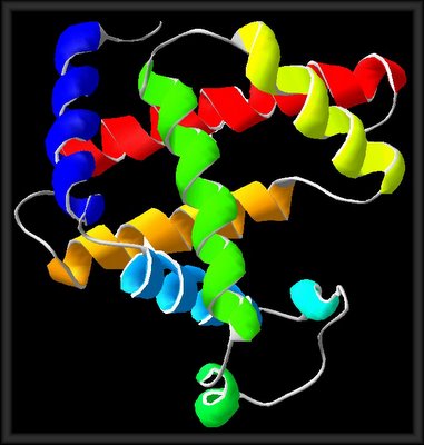
MOLECULAR BIOLOGY: ON HEDGEHOG PROTEINS: "TA "hedgehog protein" is a transmembrane protein involved in segment polarity and cell-cell signaling during embryogenesis and metamorphosis in the fruit fly Drosophila melanogaster, and in other insects and vertebrates. The expression and activity of Hedgehog proteins (Hh) exemplify a common strategy for pattern generation in metazoan embryos, namely, the specification of multiple cell fates through localized production and secretion of an instructive signal. In this manner, Hh signals regulate cell proliferation and differentiation in a diverse array of essential patterning events ranging from embryonic segmentation and appendage development in insects to neural tube differentiation in vertebrates

Study Sheds Light On Signaling Mechanism In Stem Cells, Cancer: "The primary cilium, it turns out, serves as the fulcrum in a series of acrobatic like moves between the Hedgehog signal and the Smoothened protein. Once Hedgehog has latched on to its receptor on the target cell's surface, it prompts the cell to move Smoothened, located in vesicles around the cell's nucleus, to the primary cilium. The positioning of Smoothened on the cilium, in turn, prompts downstream signaling of Hedgehog signals into the nucleus, where the instructions are issued. Just how or what the primary cilium is doing to promote Smoothened's activity is not clear, say the researchers." Hedgehog signals play an important role in prompting embryonic and adult stem cells to differentiate into some of the specialized cells that make up the body's tissues -- such as those of the brain, pancreas and skin. The scientists moved in on the role of Smoothened and the primary cilium incrementally. First, driven by their interest in Smoothened, they set out to determine where it was expressed in the embryo. They did so by developing highly specific antibodies to the protein and applying them to the tissue of an eight-day mouse embryo. The study revealed that Smoothened was modestly upregulated in cells of the node, an important early organizer tissue within the mouse embryo, and was expressed predominantly along the primary cilium of these nodal cells. This was a significant surprise. Second, to examine whether Smoothened's movement from vesicles around the nucleus to the cilium was regulated by Hedgehog signals, they carried out two studies, one involving cultured epithelial and fibroblasts cells expressing Smoothened, another involving a mouse embryo. In both cases, one set of cells was exposed to Hedgehog signals. Another set was exposed to cyclopamine, a drug that blocks Smoothened's function. In the cells exposed to the Hedgehog signals, Smoothened moved from the vesicles of the cell body to the cilium. In the cells exposed to cyclopamine, Smoothened was undetectable on the cilium. Scientists have known that cyclopamine inhibits Hedgehog signaling and can prevent Hedgehog-dependent cancers from spreading. The demonstration that the drug affected Smoothened movement to the cilium suggests how cyclopamine inhibits the Hedgehog pathway, the researchers say, and shows that the correlation between Smoothened on the cilium and pathway activation is very tight. Third, they examined whether the Smoothened protein included an amino acid sequence that other seven-transmembrane proteins require to move to the primary cilium and, if so, whether this sequence -- a so-called "motif" -- was essential to its relocation there. The answer to both questions was yes: A study of mouse cells in which Smoothened was mutated to lack the motif revealed that Smoothened no longer moved to the primary cilium. Finally, to determine Smoothened's function, they tested the mutant form of Smoothened that no longer could move to the primary cilium in epithelial cells in culture and in zebrafish embryos to see if the protein still functioned. It did not. "Thus, not only does Smoothened ciliary localization depend up on Hedgehog signaling, but Hedgehog signaling depends on a Smoothened ciliary localization motif," says Reiter. "Whether Smoothened functions at the cilium in all cell types remains to be determined. In addition, how Smoothened activates the Hedgehog pathway at the cilium remains unclear," he says. "But the current finding lays the groundwork for future studies that could ultimately have clinical benefit.""

The original news release can be found here.

histone proteins

MOLECULAR BIOLOGY: CHROMATIN DNA PACKAGING AND GENE SILENCING: "The DNA of eukaryotic genomes is packaged in nucleosomes, with approximately 167 base pairs (bp) of DNA wrapped in two left-handed turns around a core of eight histones (an [H3 4]2 tetramer and two dimers of [H2A H2B]). Histone H1 binds to the DNA where the DNA enters and exits from association with the core. 'Linker' DNA of approximately 10-50 bp extends to the next histone core. The carboxy-terminal two thirds of the core histones establish the very stable interactions that create the octamer and bind DNA to its surface, whereas the amino-terminal tails are available for interaction with other chromosomal components. The tails are substrates for a number of enzymes that modify specific amino acids of specific histones."
2006/12/15

p53

The transcription factor p53 is activated when MDM2 is inhibited by signalling by factors such as DNA damage. Once activated, p53 acts as a tumor suppressor gene by virtue of its apoptotic function. Active p53 induces the transcription of many genes, including Bax, which promotes apoptosis by stimulating the release of cytochrome c and apoptosome formation.
2006/12/13

RNA repair enzyme: T4 polynucleotide kinase

Structure and mechanism of T4 polynucleotide kinase: an RNA repair enzyme: "T4 polynucleotide kinase (Pnk), in addition to being an invaluable research tool, exemplifies a family of bifunctional enzymes with 5'-kinase and 3'-phosphatase activities that play key roles in RNA and DNA repair.

T4 Pnk is a homotetramer composed of a C-terminal phosphatase domain and an N-terminal kinase domain. The 2.0 Å crystal structure of the isolated kinase domain highlights a tunnel-like active site through the heart of the enzyme, with an entrance on the 5' OH acceptor side that can accommodate a single-stranded polynucleotide. The active site is composed of essential side chains that coordinate the beta phosphate of the NTP donor and the 3' phosphate of the 5' OH acceptor, plus a putative general acid that activates the 5' OH. The structure rationalizes the different specificities of T4 and eukaryotic Pnk and suggests a model for the assembly of the tetramer.

Li Kai Wang1, Christopher D. Lima2 and Stewart Shuman1
Structure and mechanism of T4 polynucleotide kinase: an RNA repair enzyme
The EMBO Journal (2002) 21, 3873–3880, doi: 10.1093/emboj/cdf397
Subject Categories: Structural Biology Genome Stability & Dynamics
© The content of this blog is intended for personal or educational use. All rights are reserved for commercial use of contents. All images posted on site created by site author unless otherwise stated.